Content
- A Quick Background Of X.
- Radiology Background
- History Of Medicine: Dr Roentgens Unintentional X.
- Scientists Use Ultrasounds In The 1960s.
A comprehensive history of the discovery of the X-Ray as well as the events leading up to that special occasion are given at Yale New Haven Educator's Institute. However their sacrifice some 111 years ago created a modern technology that is crucial to our health today. By January 1896 the world was gripped by "X-ray mania," as well as Roentgen was declared the discoverer of a clinical wonder. Within a year, X-rays were being used in diagnosis and also treatment and were a well established component of medicine. Then on November 8th of 1895, a German physics professor Wilhelm Conrad Roentgen made an amazing discovery. He took a tube comparable to fluorescent light bulbs, removed all the air as well as loaded it with a special gas. Public complication may develop if dental professionals remain to shield, while healthcare facilities do not.
- Today the radiologist need to follow very rigorous radiation requirements, protecting, and also radiation badge monitoring of dose as well as making use of the tiniest dosage possible to the person in achieving the answer to the medical concern.
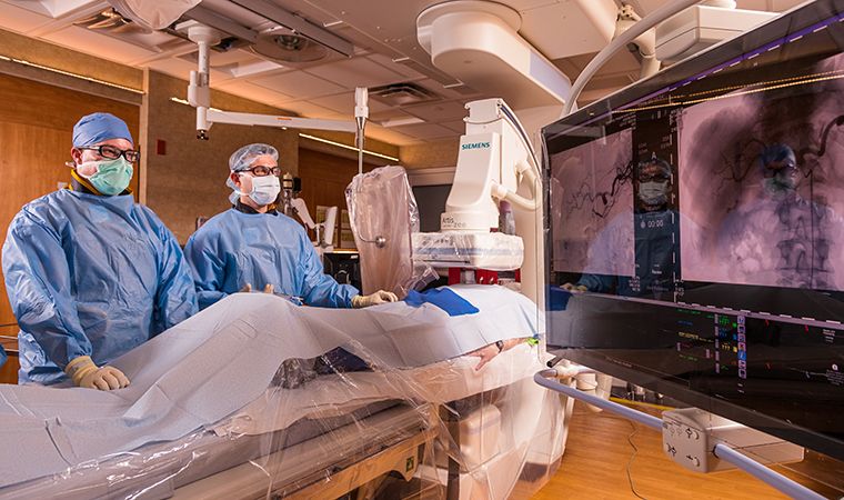
- The advancement of Nuclear Medicine in today's Radiology provides physicians the ability not only to examine anatomy, but also the physiology related to injury not available to our 1918 coworkers.
- A few hundred face, skull as well as oral film x-rays were also taken by Base Hospital # 28.
X-ray ranges can be measured either by energy dispersive or wavelength dispersive spectrometers. For x-ray diffraction applications, such as x-ray crystallography, hybrid photon counting detectors are extensively made use of. Tough X-rays can traverse reasonably thick items without being much soaked up or scattered. Consequently, X-rays are commonly made use of to image the inside of aesthetically opaque things. One of the most typically seen applications remain in medical radiography as well as flight terminal protection scanners, yet similar strategies are additionally vital in industry (e.g. industrial radiography and also commercial CT scanning) and research (e.g. tiny pet CT).
A Short Background Of X.
In 2005 SIR proposed and also ABR accepted another path called "DIRECT Pathway" to help trainees coming from other specialities learn IR; this as well was not extensively embraced. The proposition was reworked, at the very same time that total DR training was being overhauled, as well as a new proposal that would certainly bring about a double DR/IR specialization existed to the ABMS and was approved in 2012 and also became implemented in 2014. By 2016 the field had determined that the old IR fellowships would be ended by 2020. Some clinical schools in the US have actually started to incorporate a standard radiology introduction right into their core MD training. Campbell College Institution of Osteopathic Medicine additionally incorporates imaging material into their educational program early in the first year. " Board Accreditation" in diagnostic radiology calls for effective completion of 2 exams.
X-ray photons lug sufficient energy to ionize atoms and interfere with molecular bonds. This makes it a kind of ionizing radiation, as well as for that reason hazardous to living tissue. A very high radiation dosage over a short period of time causes radiation health issues, while lower doses can give a boosted risk of radiation-induced cancer cells. In clinical imaging this increased cancer risk is generally greatly surpassed by the benefits of the assessment. The ionizing ability of X-rays can be used in cancer treatment to eliminate deadly cells using radiation therapy.
Radiology Background
The term X-ray is metonymically made use of to refer to a radiographic image created utilizing this technique, in addition to the approach itself. Given that the wavelengths of difficult X-rays resemble the dimension of atoms, they are also helpful for identifying crystal structures by X-ray crystallography. By contrast, soft X-rays are quickly soaked up in air; the attenuation length of 600 eV (~ 2 nm) X-rays in water is much less than 1 micrometer. In 1914 Marie Curie created radiological autos to support soldiers hurt in World War I. The cars and trucks would certainly enable quick X-ray imaging of wounded soldiers so battlefield doctors could quickly and also a lot more precisely operate. They had to consist of a little quantity of gas as a current will certainly not flow in such a tube if they are fully evacuated. Nonetheless, as time passed, the X-rays caused the glass to take in the gas, triggering the tube to create "more difficult" X-rays till it soon stopped operating.
Who discovered the fluoroscope?
In the late 1890s, Thomas Edison began investigating materials for ability to fluoresce when X-rayed, and by https://www.washingtonpost.com/newssearch/?query=radiology the turn of the century he had invented a fluoroscope with sufficient image intensity to be commercialized.
At the transmission station, simple radiographs are gone through a digitizing equipment before transmission, while CT, MRI, ultrasound and nuclear medication scans can be sent out straight, as they are already electronic information. The computer at the getting end will certainly need to have a top quality display screen that has been checked and removed for clinical purposes. A radiologist translates clinical images on a modern image archiving and also interaction system workstation. Interventional radiology is a subspecialty of radiology in which minimally invasive treatments are done utilizing image assistance. Ordinary radiography was the only imaging modality readily available throughout the initial 50 years of radiology. As a result of its accessibility, speed, and also lower expenses compared to various other modalities, radiography is usually the first-line test of selection in radiologic diagnosis. Also despite the huge quantity of data in CT scans, MR scans and various other digital-based imaging, there are many condition entities in which the classic diagnosis is obtained by plain radiographs.
History Of Medication: Dr Roentgens Unintended X.
Thus a lot of America's radiology divisions, Yale's Department of Radiology began as a section of the Department of Surgical procedure as opposed to an independent department. Yale University College of Medication listed one radiology program for clinical students in the Division of Surgery as early as 1921, yet an official radiology residency, instead of instruction, did not begin up until the 1940s. X-Ray Day, a lot more appropriately called Imaging Day, is a short moment to appreciate the magnitude of this excellent specialty. The mammographer, another radiology professional, is reassuring the lady and taking her via the procedure of diagnosing a mass in her breast.
Phase-contrast X-ray imaging describes a selection of techniques that use phase info of a coherent X-ray beam of light to image soft tissues. It has actually come to be an essential technique for imagining mobile and histological frameworks in a variety of organic and clinical research studies. There are several technologies being made use of for X-ray phase-contrast imaging, all using various concepts to transform stage variations in the X-rays arising from a things right into intensity variations. These include propagation-based phase contrast, talbot interferometry, refraction-enhanced imaging, and also X-ray interferometry. These techniques provide higher comparison compared to normal absorption-contrast X-ray imaging, making it possible to see smaller information.
. He also collaborated with his medical college roommate, Herbert Gasser, that in 1944 got the Nobel Reward with Joseph Erlanger for gauging and identifying nerve-fiber conduction. That work had actually defied their initiatives as long as the only readily available recording gadget was the quartz fiber galvanometer, but generated when they made use of instead, the electron stream of a Brownish tube.
Do radiologists have to do surgery?
A radiologist connects your medical image to other examinations and tests, recommends further examinations or treatments, and talks with the doctor who sent you for your exam, Radiologists also treat diseases by means of https://brightonradiology.com.au/about/competitive-imaging-costs/ radiation (radiation oncology or nuclear medicine) or minimally invasive, image-guided surgery (
The radiologist or doctor after that compares the image gotten to normal anatomical photos to establish whether there is any kind of damages or blockage of the vessel. A specialized source of X-rays which is ending up being extensively utilized in study is synchrotron radiation, which is generated by bit accelerators. Its distinct attributes are X-ray results many orders of size higher than those of X-ray tubes, wide X-ray spectra, superb collimation, and also straight polarization. So, the resulting outcome of a tube contains a constant bremsstrahlung range diminishing to no at television voltage, plus numerous spikes at the characteristic lines. The voltages made use of in analysis X-ray tubes vary from about 20 kV to 150 kV and therefore the highest powers of the X-ray photons range from approximately 20 keV to 150 keV. X-rays can be generated by an X-ray tube, a vacuum tube that makes use of a high voltage to accelerate the electrons launched by a warm cathode to a high speed. The high rate electrons collide with a metal target, the anode, producing the X-rays.